Orthopaedics
Surgeons commonly operate on the bones and muscles of the body. They can transfer the location of tendons to give muscles new function, lengthen tight muscles, re-align bones, or replace whole joints with artificial devices. Computer simulation allows researchers and clinicians to answer questions like, how do muscle lengths change after treatment and how might a surgery change the forces experienced by the knee joint?
Model Surgery: Analyzing tendon transfer surgeries to restore hand function after spinal cord injury.
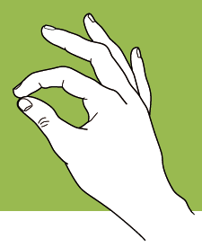 One of the key challenges for individuals who suffer from cervical spinal cord injury is regaining the ability to complete day-to-day activities independently. Hand function is compromised after this type of injury, but surgeons can perform a tendon-transfer procedure to help restore hand motions like pinching. Surgeons transfer a non-paralyzed muscle to the tendon of one of the paralyzed muscles that was responsible for the pinching function in the hand. Wendy Murray and her collaborators at Northwestern University are using a model of the hands, arms, and torso to understand how surgical technique and post-operative rehabilitation impact the success of surgery.
Murray’s team has created a model of the upper extremity that simulates the effects of tendon transfer surgery. The model includes eight muscles that individuals are still able to control after spinal cord injury. The remaining muscles in the model are “paralyzed” by setting their force-generating capacity to zero. The researchers simulate the surgery by transferring the model’s brachioradialis muscle, an elbow flexor, to the flexor pollicus longus, a muscle that flexes the thumb. Muscle activation data from electromyography, strength measurements, magnetic resonance images, and other experimental data are then used to generate a simulation of post-operative pinching that is tuned to the individual subject and the surgery they received.
The researchers have generated simulations that predict active pinch forces that match those measured experimentally after tendon transfer surgery. Their simulations show that the amount of force that subjects can generate depends heavily on how the transferred muscle is tensioned by the surgeon. Their findings also indicate that rehabilitation after surgery should focus on strengthening the elbow and wrist extensor muscles in order to maximize post-operative function. This project demonstrates how modeling and simulation can be applied to help improve surgical technique and post-operative rehabilitation, with the ultimate goal of increasing the autonomy of patients with spinal cord injury.
The following project on SimTK.org includes a model of the upper extremity like the one used to complete this work: